COVID-19 CT segmentation dataset
This is a dataset of 100 axial CT images from >40 patients with COVID-19 that were converted from openly accessible JPG images found HERE. The conversion process is described in detail in the following blogpost: Covid-19 radiology — data collection and preparation for Artificial Intelligence
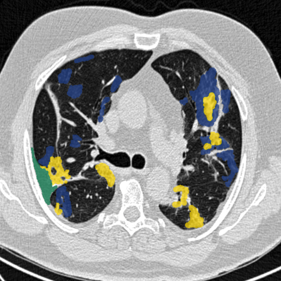
In short, the images were segmented by a radiologist using 3 labels: ground-glass (mask value =1), consolidation (=2) and pleural effusion (=3). We then trained a 2d multilabel U-Net model, which you can find and apply in MedSeg. NEW (from 2nd April 2020): Try the Beta fully automated report HERE (DICOM only).
Download data:
- Training images as .nii.gz (151.8 Mb) – 100 slices
- Training masks as .nii.gz (1.4 Mb) – 100 masks
- CSV file connecting slice no. with SIRM case no. (0.001 Mb)
- Test images as .nii.gz (14.2 Mb) – 10 slices. Kaggle competition
- This dataset on figshare (to simplify citation and ensure long-term storage and no altering of data)
An additional contribution of lung masks by Johannes Hofmanninger: Lung masks as .nii.gz (0.3Mb). His approach on Github.
Segmentation dataset nr. 2 (13th April):
We are happy to share another segmented 9 axial volumetric CTs from Radiopaedia. This dataset includes whole volumes and includes, therefore, both positive and negative slices (373 out of the total of 829 slices have been evaluated by a radiologist as positive and segmented). These volumes are converted and normalized in a similar way as above, but we have chosen not to resize these to 512×512. Download data below:
- Image volumes (308 Mb) – 9 volumes, total of >800 slices
- Covid19 masks (0.3 Mb) – includes >350 annotated slices
- This dataset on figshare
- Lung masks (1 Mb) – includes >700 annotated slices
Update 20th April: A new segmentation dataset of 20 CT scans (labels right lung, left lung and infection) is available HERE.
Comments
Ground-glass opacities have been shown to precede consolidations. Some reports have shown that early and more pronounced findings on CT correlate negatively with prognosis. Maybe volumetric measurements and ratio of ground-glass/consolidation can further enhance the prognosis estimation for COVID-19 patients. However, as radiologists, we feel it is our obligation to mention that CT should generally NOT be used for broad screening for COVID-19 as a substitute for RT-PCR.
You may use the segmentations freely, but we would very much appreciate an acknowledgment/link.